Combines 4 mm working depth with 200 micron imaging depth.
Unique Features
A compact single unit
Installed in under 20 minutes
Simple and intuitive to operation
Works in a wet lab on any flat steady surface
Works with the room lights on
Less than 100W electrical power consumption
Uniform illumination across the field of view
No edge distortions means easy tiling
Variable field of view 1.5mm to 150um without refocus or intensity adjustment
Large working distance (4mm) allows access to well plates and trans-wells
The tiled images below show many of the features of the system. The widefield image shows the captures H&E stained images. These can be
overlayed over the 2 photon images. The 2 photon images are made of 7 x 5 tiles. Each image is 650 um x 650 um making the whole tiled image
4.5 mm x 3.2 mm. The PXYL system has no image distortion, so the images can be butt coupled together with no overlap. Illumination is
flat to the edges of each image, so no processing is required. This unique feature makes tiling of large areas super easy.
PXYL system vs widefield image
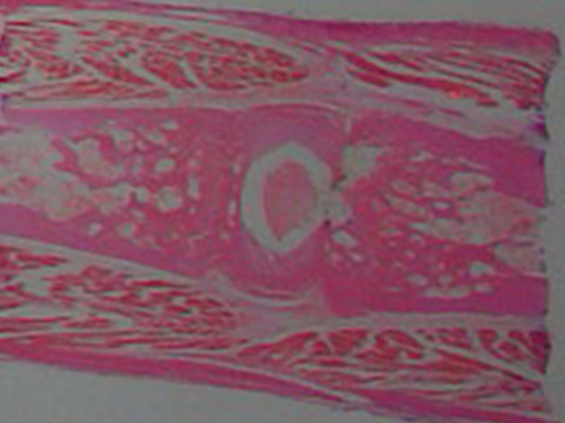
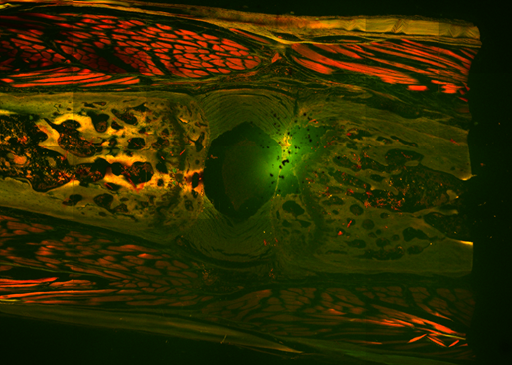
SAMPLE = Dense Connective Tissue
STAIN = Hematoxylin & Eosin (H & E)
PXYL SYSTEM vs WIDEFIELD IMAGE
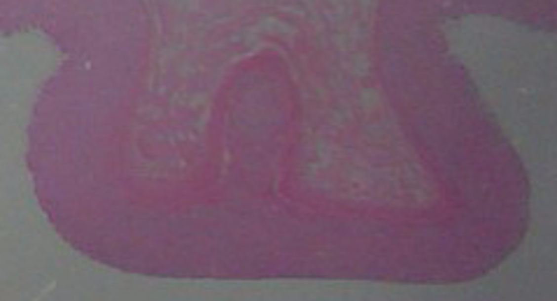
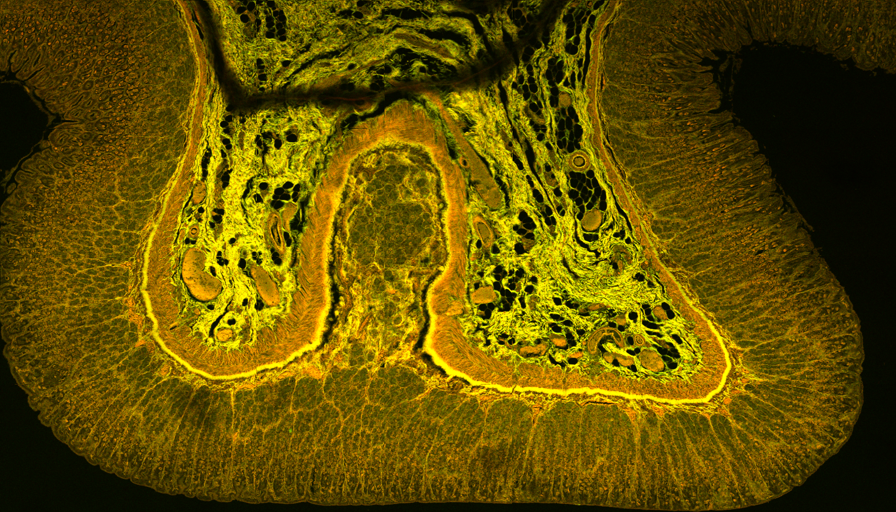
SAMPLE = Stomach Section
STAIN = Hematoxylin & Eosin (H & E)
PXYL SYSTEM vs WIDEFIELD IMAGE
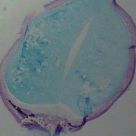
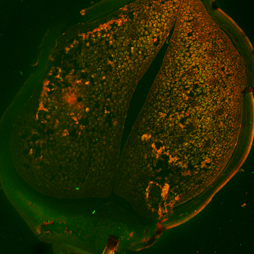
SAMPLE = Broad Bean Section
SATIN = Unstained.
Multi-photon Technology
Fully-integrated 920nm ultra-fast laser source targets GFP and RFP
Dual detector channels with customisable filters
Excellent for live studies
Very low bleaching and toxicity
1um lateral resolution
Greater than 200 micron depth penetration in 3um slices
Improve Throughput
Work close to your sample preparation area
Have your own dedicated system
Work in standard office lighting on a lab bench or table
Intuitive software for navigation, acquisition and analysis
Compatible with standard well plates and vessels
Reduce Running Costs
Hands-off, Maintenance Free System
Low power consumption (<100W ) meaning negligible heat loading
Small footprint -place anywhere
No service costs or consumables
The PXYL Advantage
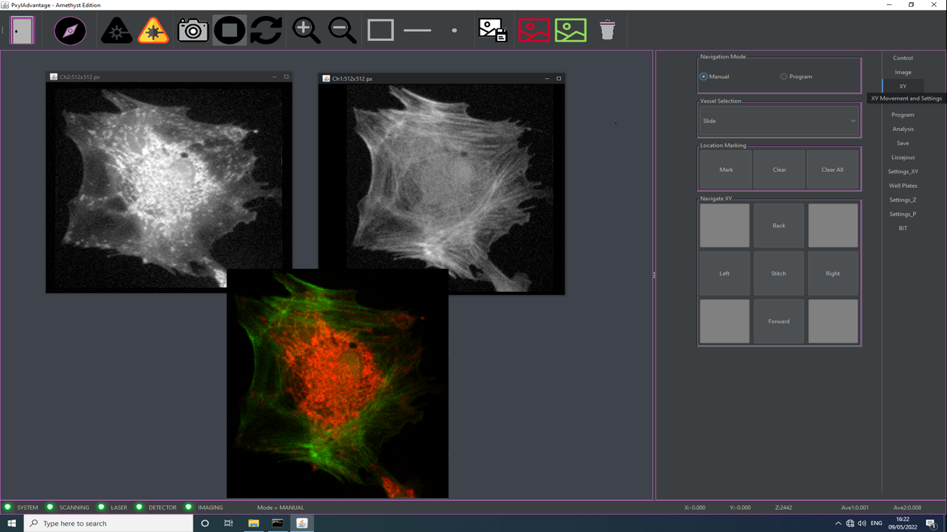
PXYL Advantage software is designed to make sample navigation simple.
Navigate your samples by pointing and clicking on the image of your sample
Effortlessly change the field of view up to 1.5 x 1.5 mms to aid navigation
Zoom in and out with no change of focus.
Auto-focus feature finds the brightest signal level in your sample
Select your vessel from a choice of well-plates, dishes and slide formats
Control laser power, detector gain and image brightness
Save images and data in a variety of standard formats (tif, png)
Demonstration
See PXYL's Table Top Multi-photon Microscope in action.
You can see a demonstration here.
Set up takes about 20 minutes from out of box to first image.
Comparison
Feature
PXYL Microscope
Typical Multi-photon
Footprint
400 x 350 mm2
2000 x 1500 mm2
Weight
25 kg
250 kg
Power Consumption
100W
4000W
Working Distance
4mm
1mm
Specification
Feature
PXYL Microscope
Detection Channels
2 independent detection channels targeting GFP and RFP
450-550nm & 575-700nm
Non-labelled second harmonic
Customised filter configurations available
Scan Size
Variable with continuous registration
60 x 60 um to 1500 x 1500 um
Pixel Density
128 x 128 px up to 1024 x 1024 px
Spatial Resolution
Transverse 1um; Axial 3um @ 920nm
Detection Mode
Sensitive non-descanned epi-fluorescence
Data Resolution
12-bit
Scan Modes
XYZ volume scanning
Uniform illumination with flat optica field to edge
Ideal for image stitiching and tiling
Auto search for signal of interest
Sample Translation
150mm x 100mm XY movement
Z-Scan Depth
4000um
Z-Scan Step increment
1um
Sample Mounting
In-vitro including standard slide, well plate or petri dish
PXYL "Advantage" Software comes as standard.
Simple controls for exploring samples in 3D.
Simple set up for automated acquistion.
Learn to use in 1 hour.
Isolation
Integrated vibration isolation system.
Applications
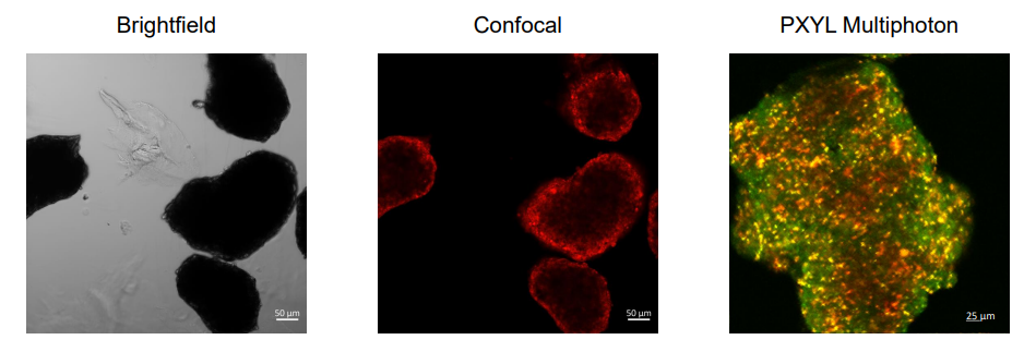
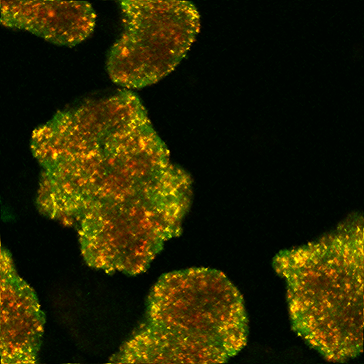
Spheroids, Organoids and Gastuloids
Whether you want to image live or fixed or cleared samples, we can help.
Mount your samples in well plates, petri-dishes or chambered slides.
Control the temperature and CO2 levels to maintain you cells over hours and days.
Take time lapse images to monitor growth and changes.
Take Z stacks or single sections, tile large areas with a couple of clicks.
Use the 4mm working distance to reach transwells with ease.
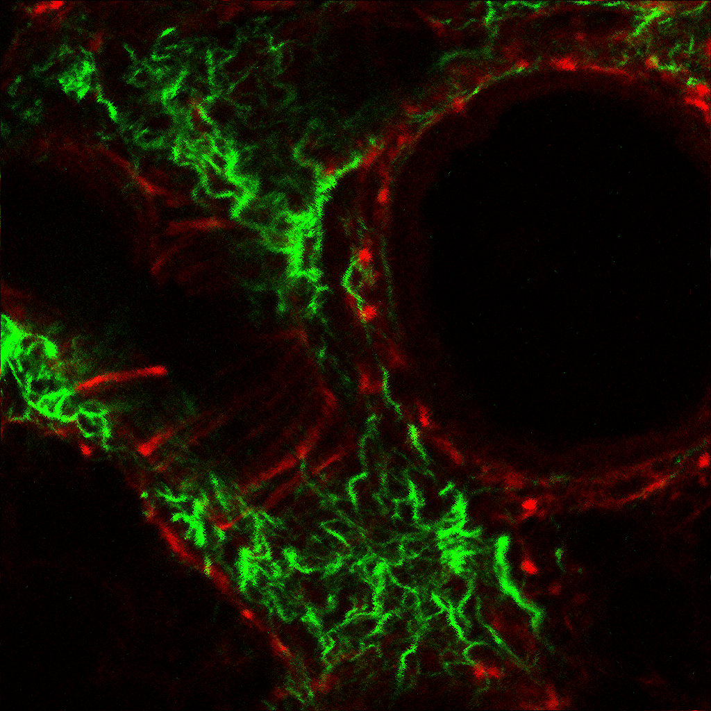
Thick Sections
The deep sectioning afforded by the longer wavelength 2-photon excitation
combined with the large working distance allows you to see through thick sections.
Like this example of a mouse lung.
The sample is 200 microns thick.
This images is taken 100um down. Field of View 300 x 300um.
PXYL system can image though the sample in 1 micron steps.
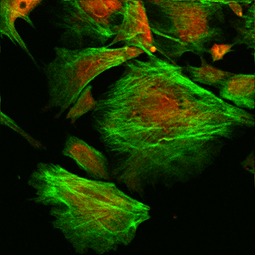
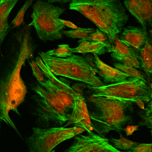
Cells
While cells are thin and can be imaged using confocal microscopes,
they are not always in a single layer and they are not always easy to reach.
PXYL's 4mm working distance allows you to search through the deepest wells.
Multi-photon also offers advantages like reduced photo-bleaching and photo-toxicity.
This means that cells can be imaged for hours without observable damage.
Ideal for longer time studies.
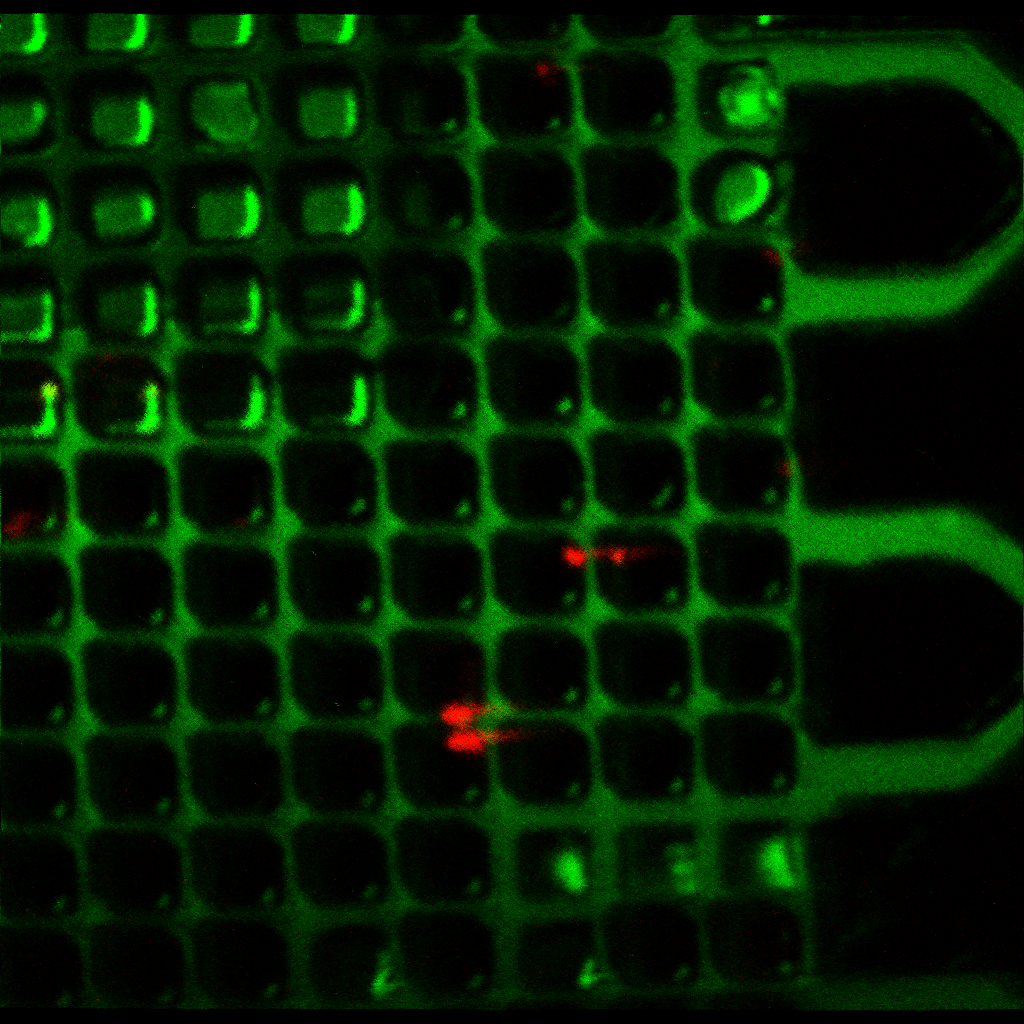
Microfluidics
Difficult to reach samples are our speciality.
Microfludic structures mounted on slide-like platforms can be hard to reach
and difficult to navigate. The 20mm FOV brightfield overview, the 1.5mm scan FOV
the 4mm working depth and long wavelength 2-photon penetration make the task easier.
PXYL's Journey
PXYL's engineers worked on some of the first multi-photon systems back in the 1990s. At the time the technique showed
great promise for deep imaging because it used longer wavelength light. Longer wavelengths are scattered less in dense
media, so they can penetrate further and suffer less optical abberations. At the time researcher were working in 2D cell cultures,
so as a technique it was largely forgotten.
Fast-forward to today and researchers are again looking for deeper and deeper imaging due to the
rise of 3D cell cultures. Multi-photon is a great choice, except for the cost, complexity, size and unkeep, Right?
Wrong! This is where PXYL comes in. We have taken our cumulated knowledge of multi-photon and
completely redesigned the system. We started with the laser and then the microscope and finally combined the two into one small box.
It really is a Black Box!
Most end users want a system that works reliably with minimal input and maximum output. That's what PXYL delivers.
PXYL's approach of redesigning the microscope and laser together has led to a much simpler
microscope with a much higher optical transmission. Typical existing microscope systems
have an optical transmission of 10-15%, while the PXYL system exhibits a transmission
of over 90%. In addition, the simpler microscope design reduces spatial and temporal
aberrations, thus delivering a high-quality optical pulse to the sample. Furthermore,
PXYL believes that table-top multi-photon microscopy with multiple fixed-wavelength
laser sources rather than one tuneable one will deliver significantly higher utility
for the end users.
Check out the Archives for more details on the PXYL origin story.
 Select your vessel from a choice of well-plates, dishes and slide formats